Incredible Examples of Electron Microscope Photography

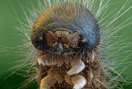
Caterpillar 30x Magnification (5mm width) | Photograph by OLIVER MECKES
Electron microscopes help bring nanoscience to life, providing a level of detail to scientists that was simply not available mere decades ago. The FEI Company is a worldwide leader in electron microscope technology. Below you will find a small collection of images from scientists around the world using FEI technology. Be sure to check out their extensive Flickr page with nearly 600 images and growing!
2. Micro-crack in Steel by Martina Dienstleder
Photograph by MARTINA DIENSTLEDER / FEI
Microcrack after bending test
Coloured by Manuel Paller
Captured by Martina Dienstleder
Instrument used: Nova DualBeam Family
Horizontal Field Width: 67µm
Voltage: 5kV
Working Distance: 6.0
Detector: ETD – SE
3. Spider’s Head by Oliver Meckes
Photograph by OLIVER MECKES / FEI
Spiders Head
Captured by Oliver Meckes
Instrument used: Quanta Family
Magnification: 50x
Vacuum: Low Vac.
Voltage: 7 kV
Spot: 3
Working Distance: app.12mm
Detector: LFD + BSE
4. Nano Mirrors on DLP Television by Regino Sandoval
Photograph by REGINO SANDOVAL / FEI
DLP Nano Mirrors
Captured by Regino Sandoval
Instrument used: Nova DualBeam Family
Magnification: 3500x
Horizontal Field Width: 73.1um
Voltage: 10kV
Spot: 5
Working Distance: 5mm
Detector: SE
5. Birth of Ladybugs by Riccardo Antonelli
Photograph by RICCARDO ANTONELLI / FEI
Birth of ladybugs
Captured by Riccardo Antonelli
Instrument used: Quanta Family
Magnification: 40x
Horizontal Field Width: 3.54 mm
Vacuum: 0.974 torr
Voltage: 10.00 kV
Spot: 5.0
Working Distance: 10.00 mm
Detector: LFD (Low vacuum)
ELECTRON MICROSCOPY
On December 29th, 1959, the noted physicist Richard Feynman issued an invitation to scientists to enter a new field of discovery with his lecture entitled “There’s Plenty of Room at the Bottom,” delivered at the annual meeting of the American Physical Society at the California Institute of Technology (Caltech). Many would credit this talk as the genesis of the modern field of nanotechnology.
Since that time there has been extraordinary progress made over that period in the field of electron microscopy, one of
the primary tools of nanoscience. Feynman called explicitly for an electron microscope 100 times more powerful than those of his day, which could only resolve features as small as about one nanometer. While we have not achieved the 100x goal – the best resolution achieved to date is 0.05 nm, a 20x improvement – FEI has indeed met his challenge to create a microscope powerful enough to see individual atoms.
For an extensive introductory overview of electron microscopy, please refer to this document.
6. Parasitic Mite on Mosquito Larva by Nicole Ottawa
Photograph by NICOLE OTTAWA / FEI
Parasitic Mite on Mosquito Larva
Captured by Nicole Ottawa
Instrument used: Quanta Family
Magnification: 200
Horizontal Field Width: app. 500 µm
Vacuum: High-Vac
Voltage: 7kv
Spot: 3
Working Distance: 9,8
Detector: LFD, BSE
7. Hydrothermal Worm by Philippe Crassous
Photograph by PHILIPPE CRASSOUS / FEI
Hydrothermal worm
Captured by Philippe Crassous
Instrument used: Quanta Family
Magnification: 57
Horizontal Field Width: 5.26 mm
Vacuum: 10-4 mbar
Voltage: 5.0
Spot: 5.0
Working Distance: 12mm
Detector: SE
8. Dehydrated Breast Cancer Cell by Wadah Mahmoud
Photograph by WADAH MAHMOUD / FEI
Breast cancer cell, fixed and dehydrated
Captured by Wadah Mahmoud
Instrument used: Inspect Family
Magnification: 5,000
Voltage: 2 kV
Spot: 2.5
Working Distance: 12.4
Detector: SE
9. Water Mite by Nicole Ottawa
Photograph by NICOLE OTTAWA / FEI
Water Mite
Captured by Nicole Ottawa
Instrument used: Quanta Family
Magnification: 700x
Horizontal Field Width: 183µm
Vacuum: 40 Pa
Voltage: 7 kV
Spot: 3
Working Distance: app. 10mm
Detector: SE+BSE
10. Corrosion on Copper Bond Pad
Photograph via FEI COMPANY
THE FEI COMPANY – ABOUT
FEI Company is the world leader in the production and distribution of electron microscopes, including scanning electron microscopes (SEM), transmission electron microscopes (TEM), DualBeam™ instruments, and focused ion beam tools (FIB), for nanoscale research, serving a broad range of customers worldwide. Nanotechnology is the science of finding, characterizing, analyzing and fabricating materials smaller than 100 nanometers (a nanometer is one billionth of a meter). FEI’s global customer base includes researchers, scientists, engineers, lab managers, and other skilled professionals.
FEI manufactures complete microscope solutions that serve the following four segments:
Research: includes a broad range of institutes, universities, and national laboratories conducting nanoscale research for a wide variety of applications including 3D nano-characterization, in situ nanoprocesses, and 3D nanoprototyping.
Natural Resources: serving the micro-analysis needs of natural resources companies focused on mining, oil & gas exploration, and geosciences. Also provides solutions for forensics including gunshot residue analysis (GSR) and forensic science. (link will redirect to our Natural Resources micro-site, fei-natural-resources.com).
Electronics: developers and manufacturers in the semiconductor, data storage and related fields with an application focus in circuit edit, 3D metrology, defect analysis, failure analysis and TEM sample preparation.
Life Sciences: includes institutes, universities, pharmaceutical companies and hospitals working in life sciences research and development in the areas of structural biology, cellular biology, tissue biology, and biomaterials.
FEI’s market-leading instruments include the latest in ion- and electron-beam technologies. From the most powerful, commercially-available microscope, the Titan™ 60-300 S/TEM, to the Magellan™, the first extreme high-resolution (XHR) scanning electron microscope, FEI produces cutting-edge tools that are revolutionizing nanoscale exploration from the classroom to the laboratory to the clean room. With a global commitment to customers before and after the sale, FEI is bringing the nanoscale within the grasp of leading researchers and manufacturers, and helping them turn some of the biggest ideas of this century into reality. FEI maintains research and development centers in North America, Europe, and Asia, and sales and service operations in more than 50 countries around the world. [Source]
11. Mosquito Larva and Parasite by Nicole Ottawa
Photograph by NICOLE OTTAWA / FEI
Mosquito Larva and Parasite
Captured by Nicole Ottawa
Instrument used: Quanta Family
Magnification: 60
Horizontal Field Width: 2000 µm
Vacuum: High-Vac
Voltage: 7kv
Spot: 3
Working Distance: 10,3
Detector: LFD, BSE
12. Iron Oxide by Francisco Rangel
Photograph by FRANCISCO RANGEL / FEI
Iron oxide
Captured by FRANCISCO RANGEL
Instrument used: Quanta Family
Magnification: 3963X
Horizontal Field Width: 75,3 ?m
Vacuum: 9.27e-7 mbar
Voltage: 20 kV
Spot: 2.0
Working Distance: 10.8
Detector: Mix: SE + BSE
13. Worm Polychaete by Philippe Crassous
Photograph by PHILIPPE CRASSOUS / FEI
Worm polychaete
Captured by Philippe Crassous
Instrument used: Quanta Family
Magnification: 150
Horizontal Field Width: 1.99mm
Vacuum: 10-4mbar
Voltage: 5
Spot: 4
Working Distance: 11.4
Detector: SE
14. Fly by Ivan Jimenez Boone
Photograph by IVAN JIMENEZ BOONE / FEI
Dirty fly
Captured by Ivan Jimenez Boone
Instrument used: MLA
Magnification: 100x
Voltage: 15kV
Spot: 7.2
Working Distance: 9.6
Detector: SE
15. Sugar Crystal by David McCarthy
Photograph by DAVID MCCARTHY / FEI
Sugar Crystal
Captured by David McCarthy
Instrument used: Quanta Family
Magnification: 187x
Horizontal Field Width: 1.37mm
Vacuum: 1.42e-4 Pas
Voltage: 0.5KV
Spot: 3.0
Working Distance: 8.5mm
Detector: SE
VISIT FEI.com FOR MORE INFORMATION
If you enjoyed this post, the Sifter highly recommends:
Alcoholic Art: Liquor Under a Microscope