20 Award-Winning Microscope Images
Olympus America Inc. is in the ninth year of sponsoring the BioScapes International Digital Imaging Competition. It honours the world’s most extraordinary microscope images of life science subjects.
The Olympus BioScapes Digital Imaging Competition recognizes outstanding images of life science specimens captured through light microscopes, using any magnification, any illumination technique and any brand of equipment. Each person entering can submit up to five movies, images, or image sequences (such as time lapse series). Entries must include information on the importance or “story” behind the images.
Below you will find a gallery of 20 winners and honourable mentions from last year’s competition. The entry deadline for this year is September 30, 2012. Be sure to visit www.olympusbioscapes.com for a complete gallery of winners, prizes and information for this year’s competition. Enjoy!
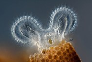
1. Rotifer Floscularia ringens feeding

FIRST PLACE: Rotifer Floscularia ringens feeding. Its rapidly beating cilia (hair-like structures) bring water that contains food to the rotifer. The “wheel animacules” were first described by Leeuwenhoek (ca.1702); when their cilia beat, they look like they have two wheels spinning on top. They live in reddish-brown tubes made of spherical “bricks.” Differential interference contrast microscopy. Charles Krebs, Issaquah, WA, USA. 2011 Olympus BioScapes Digital Imaging Competition
2. Live green brain coral (Goniastrea sp.), under water
FIFTH PLACE: Live green brain coral (Goniastrea sp.), under water. One full polyp in the center is shown with four surrounding polyps. Walled corallites are purple. All color is the natural autofluorescence of the coral with the exception of the purple, which is near-violet LED illumination to highlight neartransparent tissue. James Nicholson, NOAA/NOS/NCCOS Center for Coastal Environmental Health & Biomolecular Research, Fort Johnson Marine Lab, Charleston, SC, USA. 2011 Olympus BioScapes Digital Imaging Competition
3. Stink bug eggs
SIXTH PLACE: Stink bug eggs. Stink bugs are agricultural pests that exist throughout the world. When disturbed, they emit a characteristic foul-smelling odor. Brightfield illumination. Haris Antonopoulos, Athens, Greece. 2011 Olympus BioScapes Digital Imaging Competition
4. Fruitfly ovaries and uterus
SEVENTH PLACE: Fruitfly ovaries and uterus. The muscular and neural structure of the Drosophila melanogaster reproductive system is shown using fluorescence microscopy. The background staining of the eggs in red is a specific function of the mutant fly strain that is pictured here. Gunnar Newquist, University of Nevada, Reno, USA. 2011 Olympus BioScapes Digital Imaging Competition
5. Living diatom Mediopyxis helysia
NINTH PLACE: Living diatom Mediopyxis helysia, showing the cell nuclei and golden chloroplasts, captured using brightfield microscopy. On top there is a bacteria colony in mucilage. Specimen is from the North Sea. Wolfgang Bettighofer, Kiel, Germany. 2011 Olympus BioScapes Digital Imaging Competition
6. Spherical colonies of Nostoc commune
TENTH PLACE: Spherical colonies of Nostoc commune, a bluegreen alga. Darkfield illumination. Gerd Guenther, Duesseldorf, Germany. 2011 Olympus BioScapes Digital Imaging Competition
7. Trout alevin
Trout alevin (alevin is the second of four stages in the life cycle of a trout, when eggs hatch and the tiny fish begin to emerge). Stereomicroscopy. Robert Berdan, Calgary, Alberta, Canada. Honorable Mention, 2011 Olympus BioScapes Digital Imaging Competition
8. Weevil Eupholus, dried thorax scales
Weevil Eupholus, dried thorax scales, stack of 80 images. Darkfield. Douglas Clark, San Francisco, CA, USA. Honorable Mention, 2011 Olympus BioScapes Digital Imaging Competition
9. Hydroid collected from kelp sample
Hydroid collected from kelp sample. Epi-illumination, image stack. Mike Crutchley, Pembrokeshire, Wales, UK. Honorable Mention, 2011 Olympus BioScapes Digital Imaging Competition
10. Tintinnid ciliate of the marine plankton, Petalotricha ampulla
Tintinnid ciliate of the marine plankton, Petalotricha ampulla. This preserved specimen (Lugol’s fixed) was caught with its cilia fully extended. It is about 100 microns long. Differential interference contrast microscopy. John Dolan, Station Zoologique B.P. 28, Villefranche-sur-Mer, France. Honorable Mention, 2011 Olympus BioScapes Digital Imaging Competition
11. Two damsel bugs (Nabis sp.)
Two damsel bugs (Nabis sp.) seemingly feeding on an aphid. Background is dried leaf of Norway maple (Acer platanoides). Focus stack of 120 images. Geir Drange, Asker, Norway. Honorable Mention, 2011 Olympus BioScapes Digital Imaging Competition
12. Protozoan Elphidium crispum
Protozoan Elphidium crispum found growing on the Dorset coast of England. Brightfield. Michael Gibson, Northampton, UK. Honorable Mention, 2011 Olympus BioScapes Digital Imaging Competition
13. Juvenile live bay scallop Argopecten irradians
Juvenile live bay scallop Argopecten irradians. Through research, scientists are trying to help restore scallop populations in Rhode Island. Kathryn Markey, Aquatic Diagnostic Laboratory, Roger Williams University, Bristol, RI, USA. Honorable Mention, 2011 Olympus BioScapes Digital Imaging Competition
14. Cross-section of bulrush (Juncus sp.)
Cross-section of bulrush (Juncus sp.) leaf, autofluorescing red (chlorophyll on external side of leaf) and blue (vascular bundles). Diameter of the stalk is approximately 3mm. Jan Martinek, Ostrov, Czech Republic. Honorable Mention, 2011 Olympus BioScapes Digital Imaging Competition
15. Young sporangia of slime mold Arcyria stipata
Young sporangia of slime mold Arcyria stipata. Fluorescence. Dalibor Matýsek, Mining University – Technical University of Ostrava, Ostrava, Czech Republic. Honorable Mention, 2011 Olympus BioScapes Digital Imaging Competition
16. Sporangium of the slime mold Craterium minutum
Sporangium of the slime mold Craterium minutum. Fluorescence. Dalibor Matýsek, Mining University – Technical University of Ostrava, Ostrava, Czech Republic. Honorable Mention, 2011 Olympus BioScapes Digital Imaging Competition
17. Sporangium of the slime mold Physarum leucophaeum
Sporangium of the slime mold Physarum leucophaeum. Fluorescence. Dalibor Matýsek, Mining University – Technical University of Ostrava, Ostrava, Czech Republic. Honorable Mention, 2011 Olympus BioScapes Digital Imaging Competition
18. Eye of a damselfly
Eye of a damselfly. The image reveals the regular, crystal-like hexagonal lattice of the eye’s elements. Projection of confocal stack, 20x objective. Igor Siwanowicz, Max Planck Institute for Neurobiology, Munich, Germany. Honorable Mention, 2011 Olympus BioScapes Digital Imaging Competition
19. Detail of a pod of flowering legume Scorpius muricatus
Detail of a pod of flowering legume Scorpius muricatus. Stereomicroscopy, darkfield illumination. Viktor Sýkora, Hyskov, Czech Republic. Honorable Mention, 2011 Olympus BioScapes Digital Imaging Competition
20. Cat tooth in cross section
Cat tooth in cross section, showing the membrane surrounding the outside of the tooth. Darkfield illumination. Image composed of 38 images. Tim Tiebout, Rochester Institute of Technology, Rochester, NY, USA. Honorable Mention, 2011 Olympus BioScapes Digital Imaging Competition
If you enjoyed this post, the Sifter highly recommends:


Sign up to get our BEST stories of the week straight to your inbox.