Innovation In Ultrasound Technology Allows Doctors To Get Incredibly Precise Images Of Internal Organs In 4D Without Surgery

Shutterstock
When doctors are trying to get a look inside the body to try to learn something about a patient, they often have to weigh out several different options to decide which one is best.
Cutting someone open is the best way to get a really good up close look, but it has a lot of risk to it, so it shouldn’t be done unless absolutely necessary. Another option is to use an MRI, which is less invasive, but it can be hard on the patient if they are claustrophobic. It also has some limitations on what the images can show.
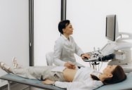
For a long time, the safest and easiest way to take a look inside someone’s body has been the ultrasound. Ultrasounds are so safe that they are the standard option for looking at a child within a pregnant mother.
When first developed, the images returned from an ultrasound were pretty low-resolution, and it took a real expert to figure out what they were looking at. Years ago, improved technology introduced ‘3D ultrasounds’ that could show depth and even some level of color, which was absolutely amazing.
Even with that 3D technology, however, the images were limited in what they could show when you get down to the very small levels. For example, if a doctor were trying to diagnose a vascular issue where the veins can be less than 100 micrometers, it is essentially useless.
A team of scientists in France, however, may have solved this problem. They have developed a 4D ultrasound that is proving that it can generate an extremely precise image for doctors to use. In a statement about the tool, senior author Clement Papadacci, said:
“The originality of these results lies in the fact that these images allow us to visualise the vessels of an entire organ at very small scales (less than 100 micrometres) – this 4D image resolution is unprecedented, as is the ability to observe an entire large organ and its flow dynamics.”
You can see the technology in action in this quick video:
This will provide doctors with a level of detail not previously possible without cutting into the patient (and even then, it wasn’t always visible). Papadacci explains:
“Used in clinical settings, this new technology could become a major tool for better understanding vascular dynamics as a whole, from the largest vessels to the pre-capillary arterioles. It could also help advance the diagnosis of microcirculation disorders and the monitoring of treatments for small vessel diseases, which are complex to diagnose and are diagnosed by ruling out other pathologies.”
So far, this innovation has been tested successfully using flow patterns in plastic tubing, ex vivo experiments on the hearts of pigs, in vivo scans on live pigs, and more. The results were extremely good, offering a complete visualization of the vascular network.
Shutterstock
The next stage of their trials is to begin using them on humans. Since this is an advancement on an existing technology, they can often get trials approved more quickly than would be possible with brand new inventions or medications. The information about this system was published in a study in Nature Communications.
If they get positive results on the human trials, this new 4D ultrasound technology could be rolled out into clinical environments soon.
If you enjoyed that story, check out what happened when a guy gave ChatGPT $100 to make as money as possible, and it turned out exactly how you would expect.


Sign up to get our BEST stories of the week straight to your inbox.